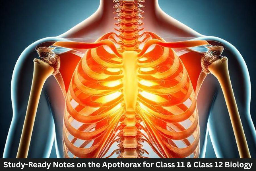
The apothorax refers to the chest region of the human body that houses some of the most essential organs responsible for keeping us alive. In classical anatomical terminology, this word described the upper trunk area enclosed by the ribs, where the heart and lungs function together to maintain breathing and circulation. In modern anatomy, the more commonly used term is thorax, which refers to the same chest cavity between the neck and the abdomen.
Think of the apothorax as the body’s central control room for life-sustaining systems. Every breath you take and every heartbeat you feel originates in this protected chamber. The lungs bring oxygen into the body, while the heart pumps oxygen-rich blood throughout the body. Without the apothorax providing a stable environment for these organs, these essential functions would not occur efficiently.
The apothorax is also remarkable because it is both strong and flexible. The rib cage shields vital organs from injury while still allowing expansion and contraction during breathing. Each inhalation slightly expands the chest cavity, allowing the lungs to inflate like balloons, and each exhalation gently compresses the space to push air back out.
This balance of protection and flexibility highlights why the apothorax plays such an important role in the human body. It is not simply an empty space in the torso—it is an intricate anatomical region where several systems interact continuously to sustain life.
Historical Origins of the Term Apothorax
The term apothorax has interesting linguistic roots that date back to earlier anatomical studies. It originates from Greek words: “apo,” meaning away from or upper, and “thorax,” meaning chest. Together, the word historically referred to the upper chest region or the area surrounding the thoracic cavity.
In early anatomy texts, scientists and physicians often used a wide variety of terms to describe body structures. Standardized terminology had not yet been established, which meant that the same region of the body might be described in different ways by different authors. As a result, the word apothorax sometimes appeared in classical medical writings to describe the chest area or supportive structures around it.
Over time, anatomy became more standardized. Global medical communities agreed to use clearer and more consistent terminology to prevent confusion in research and clinical practice. This led to the adoption of the term thorax as the official name for the chest cavity.
Even though the word apothorax is rarely used today, it still appears in educational materials and historical discussions of anatomy. Understanding it helps students connect older anatomical language with modern terminology. In many ways, learning about the apothorax provides insight into how medical science has evolved and how anatomical knowledge has become more precise over the centuries.
Where Is the Apothorax Located in the Human Body
Position Between the Neck and Abdomen
The apothorax occupies a central location in the human body. It lies between the neck (cervical region) and the abdomen, forming the main chest cavity where several vital organs are located.
If you place your hand over the middle of your chest and take a deep breath, you are essentially touching the outer region of the apothorax. Beneath that surface lies a complex structure made up of bones, muscles, connective tissues, and internal organs.
This region is especially important because it connects several major body systems. The respiratory system, circulatory system, and portions of the digestive system all pass through or function within this space. The trachea carries air to the lungs, blood vessels transport blood from the heart to the body, and the esophagus passes through the chest on its way to the stomach.
Another fascinating feature of the apothorax is how it allows constant motion without compromising protection. Each breath you take causes the rib cage to move slightly outward and upward. This movement increases the volume inside the chest cavity, allowing the lungs to expand and draw air into the body.
Because of its central location and its involvement in essential bodily processes, the apothorax acts as a bridge between different regions of the body. It links the head and neck above with the abdomen below while supporting several organs that keep the body functioning every second of the day.
Anatomical Boundaries of the Apothorax
The apothorax is defined by several anatomical boundaries that create a protective chamber for internal organs. These boundaries form a three-dimensional enclosure that allows movement while maintaining structural stability.
At the top of the apothorax, the boundary is known as the thoracic inlet. This opening connects the chest cavity to the neck and allows important structures like the trachea, esophagus, and major blood vessels to pass through. It acts like a gateway between the upper body and the chest.
The lower boundary is formed by the diaphragm, a dome-shaped muscle that separates the chest cavity from the abdominal cavity. This muscle plays a crucial role in breathing by contracting and relaxing to control the movement of air in and out of the lungs.
The sides of the apothorax are enclosed by the rib cage and intercostal muscles. These structures provide both strength and flexibility, allowing the chest to expand during inhalation and contract during exhalation.
Finally, the front and back boundaries consist of the sternum (breastbone) at the front and the thoracic vertebrae of the spine at the back. Together, these bones create a sturdy yet movable framework that protects the delicate organs within the chest cavity.
Structure of the Apothorax
The Rib Cage and Thoracic Skeleton
The structural framework of the apothorax is built around the rib cage, which serves as the main protective barrier for the organs inside the chest. The rib cage consists of 12 pairs of ribs, the sternum in the front, and the thoracic vertebrae in the back. These bones work together to form a strong but flexible cage around the lungs and heart.
The ribs are arranged in a curved pattern that creates space for the lungs to expand during breathing. Each rib connects to the spine at the back and most connect to the sternum through cartilage at the front. This design allows the rib cage to move slightly with each breath, expanding during inhalation and contracting during exhalation.
This skeletal framework is incredibly efficient. It provides strong protection against physical impacts while still allowing enough flexibility for constant movement. Without this balance of strength and mobility, breathing would become difficult and the organs inside the chest would be more vulnerable to injury.
The rib cage also supports several muscles that assist with breathing. These muscles, known as intercostal muscles, sit between the ribs and help control the expansion and contraction of the chest cavity. When they contract, the ribs move outward, increasing the volume inside the chest and allowing the lungs to fill with air.
Because of this sophisticated design, the apothorax is able to protect vital organs while also supporting one of the most important functions of life—breathing.
Muscles and Soft Tissue Surrounding the Apothorax
Beyond the skeletal framework, the apothorax is surrounded by a network of muscles, connective tissues, and membranes that help maintain its structure and function. These soft tissues play a crucial role in breathing, posture, and protection of internal organs.
One of the most important muscle groups in this region is the intercostal muscles, which run between the ribs. These muscles assist the rib cage in expanding and contracting during breathing. When they contract, the chest cavity becomes larger, allowing air to flow into the lungs. When they relax, the chest cavity shrinks and air is pushed out.
The chest wall also includes layers of connective tissue and fascia that stabilize the rib cage and anchor muscles to the skeleton. These tissues help maintain the shape of the chest and distribute mechanical forces during movement.
Additionally, the lungs are surrounded by a thin membrane called the pleura, which allows them to move smoothly against the chest wall as they expand and contract. Without this lubricated surface, breathing would create friction and discomfort.
Together, these muscles and tissues transform the apothorax into a dynamic structure that can move constantly without compromising stability or protection.
Major Organs Located in the Apothorax
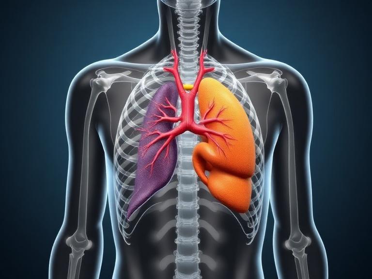
The Heart
The heart is one of the most vital organs located within the apothorax. Positioned slightly left of the center of the chest, this muscular organ acts as the body’s primary pump for circulating blood. Every minute, the heart pushes oxygen-rich blood through arteries to reach tissues and organs throughout the body.
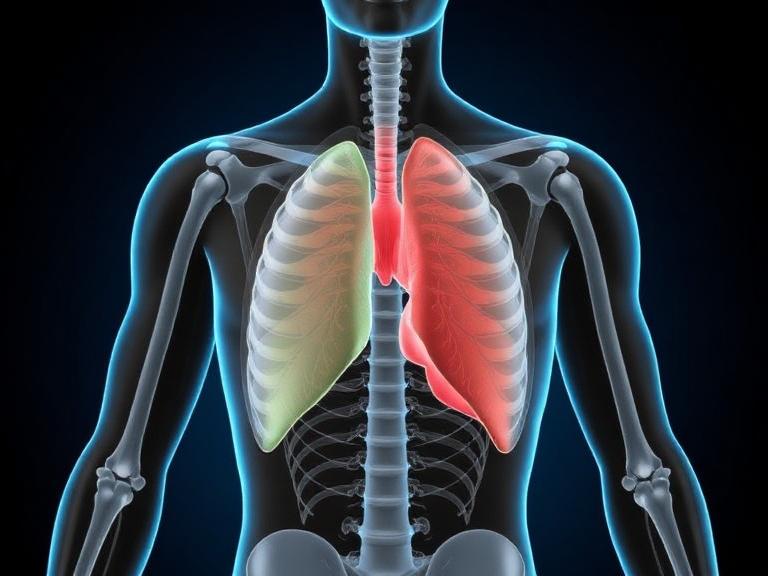
The Lungs
The lungs occupy the largest space inside the apothorax. These two spongy organs are responsible for gas exchange, a process that allows oxygen to enter the bloodstream while carbon dioxide leaves the body.
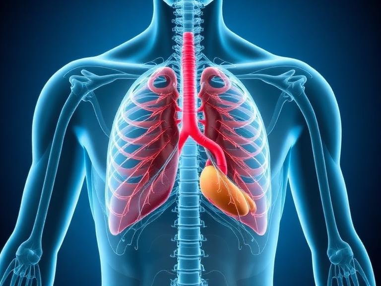
Trachea and Bronchi
The trachea, often called the windpipe, is the airway that connects the throat to the lungs. Inside the apothorax, the trachea divides into two bronchi, which lead into each lung and allow air to travel deeper into the respiratory system.
Major Blood Vessels
Several major blood vessels pass through the apothorax, including the aorta, vena cava, and pulmonary arteries and veins. These vessels transport blood between the heart, lungs, and the rest of the body, forming a critical part of the circulatory system.
Functions of the Apothorax
Role in Breathing and Oxygen Exchange
The apothorax plays a central role in breathing. As the rib cage expands and the diaphragm contracts, the chest cavity increases in volume. This creates negative pressure that pulls air into the lungs.
Role in Circulation
The heart’s location within the apothorax allows it to efficiently pump blood through major arteries and veins that branch throughout the body.
Protective Function for Vital Organs
The rib cage and surrounding muscles act as a protective barrier, shielding the heart and lungs from injury while allowing necessary movement.
The Diaphragm and Its Relationship With the Apothorax
How the Diaphragm Controls Breathing
The diaphragm forms the floor of the apothorax and acts as the primary muscle responsible for breathing. When it contracts, it flattens downward and increases the volume of the chest cavity, allowing air to enter the lungs.
Apothorax vs Thorax: Understanding the Difference
| Feature | Apothorax | Thorax |
|---|---|---|
| Usage | Historical term | Modern anatomical term |
| Meaning | Upper chest region | Chest cavity |
| Medical usage | Rare | Standard in medicine |
Both terms refer to the same region of the body, but thorax is now the widely accepted scientific term.
Common Medical Conditions Affecting the Apothorax
Several medical conditions can affect the organs inside the apothorax, including respiratory infections, heart disease, and lung disorders. Injuries such as rib fractures or collapsed lungs can also impact this region and interfere with breathing.
Importance of the Apothorax in Human Health
The apothorax supports some of the most essential biological processes. Every breath you take and every heartbeat you feel depends on the structures contained within this region. Because it houses both the lungs and the heart, any problem affecting the apothorax can quickly influence the entire body.
Conclusion
The apothorax represents one of the most important anatomical regions of the human body. Located between the neck and abdomen, it forms the chest cavity that protects and supports the heart, lungs, and major blood vessels. Even though modern anatomy typically uses the term thorax, understanding the concept of the apothorax helps explain how the body organizes and protects its most vital organs.
From breathing and circulation to structural protection, the apothorax plays a critical role in maintaining life. Every heartbeat and every breath occurs within this carefully designed cavity. Learning about its structure, organs, and functions gives us a deeper appreciation for the remarkable complexity of the human body.
FAQs
1. What is the apothorax in simple terms?
The apothorax refers to the chest cavity that contains vital organs such as the heart and lungs.
2. Is apothorax the same as thorax?
Yes. Apothorax is an older anatomical term that refers to the same chest region now commonly called the thorax.
3. What organs are found in the apothorax?
The heart, lungs, trachea, bronchi, and major blood vessels are located in the apothorax.
4. What protects the organs in the apothorax?
The rib cage, sternum, spine, and surrounding muscles protect the organs inside the chest cavity.
5. Why is the apothorax important?
The apothorax houses organs responsible for breathing and blood circulation, making it essential for survival.